
Procedures
What is an "FA"
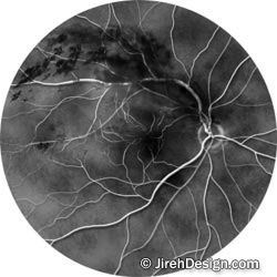
Fluorescein Angiography (FA) - A fluorescein angiogram (fluorescein - the type of dye that is used; angiogram - a study of the blood vessels - in this case, on your retina) is a valuable test that provides information about the condition of the retina. FAs are useful for evaluating many eye diseases that affect the retina in the back of the eye.
The study is performed by injecting a sodium-based dye into an arm vein. The dye appears in the blood vessels on the retina in 10-15 seconds on average. As the dye travels through the retinal blood vessels, an ophthalmic photographer shoots pictures of the retina with a special retinal camera. If there are any abnormalities on the retina, the dye will usually reveal them by leaking, staining or by its inability to get through blocked blood vessels.

Although statistically very rare, adverse reactions to the intravenous dye have been reported. You should discuss the risks and benefits of this common study with your ophthalmologist.
At Carolina Macula and Retina, these photos are taken with a digital camera system, allowing Dr. Gross to interpret the results immediately.